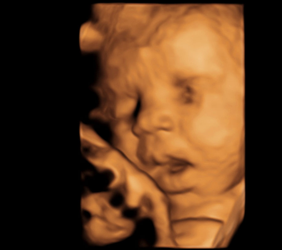

Scheduling an ultrasound appointment with Baby in Sight 3D is quick and easy. In our case those sonogram images can be in 2D, 3D or HD Live. Now that we’ve addressed what an ultrasound is, how does that differ from a sonogram?Ī sonogram is simply the resulting ultrasound image or picture generated by the ultrasound machine.

In addition, our GE ultrasound equipment is outfitted with the latest advancement in 4D imaging called HD Live (5D) enabling us to view baby with unparalleled clarity and realism. A pioneer in the field, Brown played an important role in the history of ultrasound. The 3-D Multiplanar scanner was developed through the work of Thomas Graham Brown, who invented 3D Multiplanar scanner and brought it to market in 1975.
#3D SONOGRAM SOFTWARE#
Our high-end obstetrical ultrasound system has additional specialized software that can transform the standard 2D black and white ultrasound image into a volume or 3-dimensional image. Three-dimensional ultrasound and its use in medicine is a relatively new addition to a physician’s toolkit. This resulting image looks more like what youre used. Sound waves reflect back to the wand from internal structures and are interpreted by sophisticated system software creating an ultrasound image. In a 3D ultrasound, many 2D images are taken from various angles and put together to form a 3D image. Mothers are generally most excited to see 3D images of their babys. 3-D/4-D ultrasound to obtain keepsake fetal portraits is an elective, nonmedically necessary procedure which both the FDA and the American Institute of Ultrasound in Medicine discourage due to the lack of information about long-term effects of ultrasound exposure. Book an appointment today and find 3d 4d ultrasound prices near me. 1414A Newkirk Avenue, Brooklyn, NY 11226. So then what is the difference between an ultrasound and a sonogram?Īn ultrasound involves the utilization of high frequency sound waves ( not ionizing radiation as some think) emitted into the body by a hand-held device coated with hypoallergenic water-based gel. 3D ultrasounds may be utilized to better visualize certain anatomical parts of your baby. Get the newest of our beautiful first baby pictures through 3d ultrasound Dallas TX. Sharmeen Sultana MD PLLC is the ideal decision to get benefit from doing a 3d/4d sonogram brooklyn since it is easy, has no experience with radiation, has clear pictures, and is low cost. You’ve likely heard your doctor or a television personality order a sonogram or an ultrasound. Often the medical community uses the terms sonogram and ultrasound interchangeably and it is widely believed they are synonymous. 3d Sonogram w 4d HD Ultrasound Sessions Featured Products On Sale 'Prince' Or 'Princess' Gender Ultrasound Determination Reveal Sonogram 129.00 89.00 GIFT CARDS Great for 3d 4d ultrasounds, Gender reveal sonograms, Maternity & Newborn Photography Sessions, Bridal Shower gift, Dallas, Fort Worth 50. Official statement.Are you looking for a 3D sonogram of your Baby? doi:10.1016/j.ultrasmedbio.2014.12.677Īmerican Institute for Ultrasound in Medicine.

More expecting parents than ever are paying to get photos and videos of their babies that are more lifelike than the 2-D ultrasounds from their doctor’s offices. A review on real-time 3D ultrasound imaging technology. A quick Google search reveals that nearly a dozen businesses in the Dallas-Fort Worth area offer 3-D and 4-D keepsake ultrasound services. Please call our 3D / 4D facility in Fort Worth at 81, 81 should you have any questions regarding our 3D or 4D ultrasound packages.

Sonographic markers for early diagnosis of fetal malformations. How do health care providers diagnose Down syndrome?. Ultrasound exams.Įunice Kennedy Shriver National Institute of Child Health and Human Development. A 3d ultrasound is a static 3 dimensional image of your baby and 4D adds the dimension of time, allowing you to see your babys movements in the womb in. doi:10.1002/j.Īmerican College of Obstetricians and Gynecologists. A pictorial guide for the second trimester ultrasound. Avoid fetal "keepsake" images, heartbeat monitors.īethune M, Alibrahim E, Davies B, Yong E.


 0 kommentar(er)
